The Retina

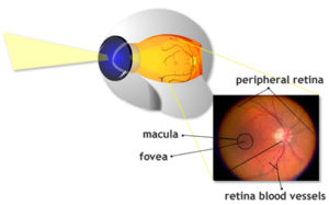
The retina is the thin transparent layer covering most of the inside lining of the eye. It is often compared to the film inside a camera because like film it is light sensitive and images are brought to focus on it. The retina consists mainly of nerve cells and is actually an extension of the brain. The retina can be broken down into 3 main regions, the macula, the fovea, and the peripheral retina or periphery. (view cross section)
Most of the specialized cone cells are concentrated in the macula and fovea located close to the center of the retina. These cells provide sharp central vision and are responsible for you seeing color. The fovea is the center of the macula. In order to have sharp detailed vision, such as reading, threading a needle, etc., it is crucial for this area of the retina to be functioning properly. If this region gets damaged enough it can leave a person with peripheral vision only.
The peripheral region constitutes the area surrounding the macula and makes up 95% of the retina. It is often referred to as your side vision. High concentrations of rod cells are found in this region and are used in low light conditions. An example would be to stare at a word on this page. Hold your hand up to either side. As you’re staring at the word you’ve chosen you’ll still be able see your hand, fingers and its general shape, but not the details of it, such as the lines and creases in it.
The head of the optic nerve is another region of the back lining of the eye. It is a natural or physiologic blind spot because no rod or cone cells are located there. When light is focused to the retina stimulating the rod and cone cells, they send an electric nerve impulse via nerve fibers in the retina. These nerve fibers converge forming the optic nerve which act as cable transmitting the signals to the visual cortex at the back of the brain. This is also where the central retina artery enters and the central retina vein exists.
